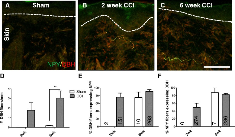
Fig. 7.

CCI induced a sprouting of sympathetic fibers into the upper dermis of the skin, and these fibers often expressed NPY. Sympathetic fibers labelled with DBH (in red), were rarely observed in the upper dermis of the skin in 6 week sham animals (a). 2 (b) and 6 (c) weeks after CCI, sympathetic fibers sprouted into the upper dermis, and many expressed NPY. The quantification of the number of DBH fibers per mm of upper dermis is shown in (d). The quantification of the percentage of DBH fibers that expressed NPY is shown in e and the values written on the graphs indicate the absolute number of sprouted sympathetic fibers counted across animals. The quantification of the percentage of NPY fibers that expressed DBH is shown in f and the values written on the graphs indicate the absolute number of NPY-positive fibers counted across animals. Dashed lines represent the border between the epidermis and the upper dermis. **p < 0.01 by 2 way ANOVA with Bonferroni post hoc (n = 6). Scale bar = 50 μm
