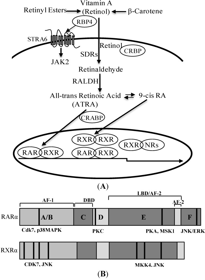
Figure 2.
Retinoid metabolism and the structure of retinoid receptors. (A) Retinol binds to RBP4 (retinol binding protein 4) in plasma. RBP4 bonds to STRA6, resulting in the activation of the JAK2 signaling pathway. Retinol bound with cellular retinol-binding protein (CRBP) is converted to retinaldehyde by short-chain dehydrogenase/reductase (SDR); and then to all-trans retinoic acid (ATRA) by retinaldehyde dehydrogenase (RALDH); (B) Schematic representation of the functional domains and the major phosphorylation sites of RARα (retinoic acid receptor α) and RXRα (retinoid X receptors α). The DNA-binding domain (DBD) and the ligand-binding domain (LBD) are schematically represented (not to scale). The functional AF-1 and AF-2 domains lie in the A/B and E regions, respectively. Phosphorylation sites are shown in a bold black line.

