
Fig. 1.
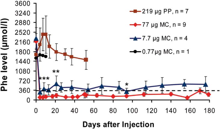
Long-term correction of hyperphenylalaninemia in PKU (Pahenu2) mice after delivery of MC-DNA vectors expressing the Pah-cDNA from the liver-specific P3 promoter. Vectors were delivered to the liver of adult PKU mice by a single HTV injection. Seven mice were injected with the parental plasmid vector pMC.PKU20 (■; 219 μg or 53.7 pmol; 3 × 10¹³ particles), and 9 mice with an equimolar amount of MC vector MC.PKU20 (◆; 77 μg or 53.7 pmol; 3 × 10¹³ particles). The minicircle vector MC.PKU20 was also used for titration studies by injecting 4 mice at a dosage of 7.7 μg (▲; 5.37 pmol or 3 × 10¹² particles) and 1 mouse with 0.77 μg (●; 0.54 pmol or 3 × 10¹¹ particles). Blood was collected on Guthrie filter cards from tail vein bleedings prior to DNA vector infusion (week 0) and periodically after vector delivery, and depicted as the mean ± SD for each treatment group. Phe concentrations were determined by tandem mass spectrometry. The broken line represents the defined therapeutic levels of blood Phe (≤360 μmol/L). Serum Phe levels in treated mice injected with 77 μg and 7.7 μg MC-DNA vectors were compared for statistically significant difference by one-tailed Student t test, ***P < 0.005 at day 4, **P < 0.05 at day 21, and *P < 0.1 at day 94 following vector administration.