
Fig. 3.
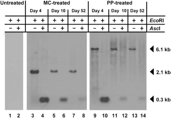
Southern analysis demonstrates that DNA vectors remain episomal and unmethylated on CpG island in the P3 promoter. Total DNA was prepared from the livers of PKU mice at a range of timepoints following hydrodynamic vector infusion. Genomic DNA was digested with EcoRI either alone or with the CpG methylation sensitive enzyme AscI. The blotted DNA was probed with 32P labeled DNA amplified from the P3 promoter, which is common to both constructs. In the single EcoRI digest, the vectors are linearized and the probe detects a vector-sized band (6.1 kb PP pMC.PKU20 or 2.1 kb MC MC.PKU20). If the DNA is not methylated, then the double digest using EcoRI and AscI removes the P3 promoter sequence and the probe detects a smaller 0.3 kb fragment. Lanes 3 to 8 represent MC-treated PKU mouse livers (77 μg), while lanes 9 to 14 represent PP-treated PKU mouse livers (219 μg). Lanes 1 and 2, PKU untreated; lanes 3, 4, 9, and 10, day 4; lanes 5, 6, 11, and 12, day 10; lanes 7, 8, 13, and 14, day 52.