Fig. 1.
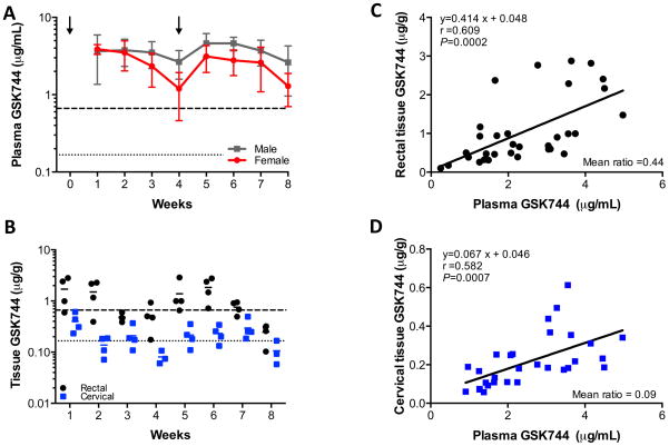
GSK744 LA plasma PK profile and tissue distribution in Depo-Provera-treated female rhesus macaques. Eight female rhesus macaques were injected IM with 30 mg Depo-Provera on weeks -3 and 2, and with 50 mg/kg of GSK744 LA on weeks 0 and 4. (A) GSK744 plasma concentrations from Depo-Provera-treated female rhesus macaques (red) were compared with male rhesus macaques (black) dosed with 50 mg/kg GSK744 LA on weeks 0 and 4. Mean ± SD are shown. Dotted and dashed horizontal lines represent 1x and 4x PAIC90, respectively. (B) Rectal and cervical tissue distribution of GSK744 was assessed from pinch biopsies each week in a subset (n=4) of Depo-Provera-treated rhesus macaques. Each symbol represents tissue concentrations from an individual macaque. Solid lines represent the mean for the group. Dotted and dashed horizontal lines represent 1x and 4x PAIC90, respectively. Correlation of (C) rectal tissue GSK744 or (D) cervical tissue GSK744 and plasma GSK744 concentrations. Each symbol represents the simultaneous plasma and tissue concentrations of an individual macaque.

