
Fig 4.
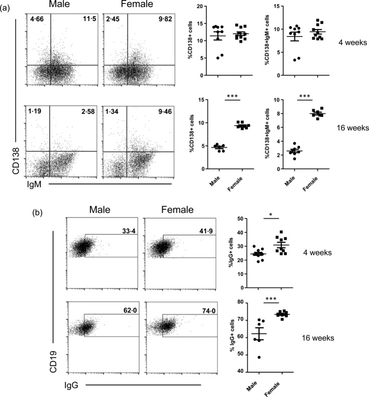
Female SWR × NZB F1 (SNF1) mice show higher numbers of plasma cells in the gut mucosa. Peyer's patches of 4- and 16-week-old male and female mice were stained for surface CD19, CD138, immunoglobulin (Ig)M and IgG and analysed by fluorescence activated cell sorter (FACS). (a) The frequencies of CD138 single-positive and CD138 and IgM double-positive plasma cells are shown. (b) CD19+ cells with surface expression of IgG are shown. Representative FACS plots (left panels) and mean ± standard error of the mean of percentage values (right panels) are shown.