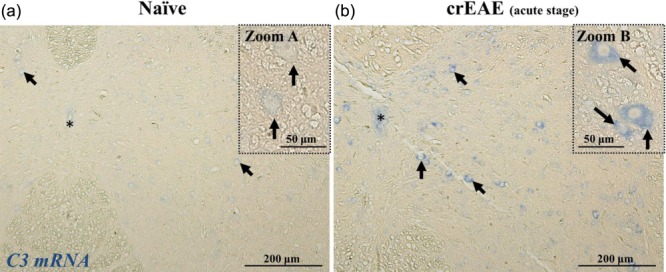
Fig 6.

C3 is produced locally by neurones in chronic relapsing experimental autoimmune encephalomyelitis (crEAE). In-situ hybridization for C3 mRNA on spinal cords from naive mice (a, representative of four mice) and crEAE mice at acute stage of disease (b, representative of four mice), showing the C3 mRNA signal (in blue) in neurones (arrows and zoom), as inferred by the morphology and large nuclei. Note the light blue staining in neurones of naive mouse spinal cord (zoom A), whereas a strong blue staining is detected in the cytoplasm of neurones in the spinal cord of crEAE mice at acute stage of disease (zoom B). Images are taken at equivalent spinal cord locations of naive and crEAE mice. The central canal is indicated by the asterisk (*). Scale bar A and B, 200 μm; zoom A and B, 50 μm.
