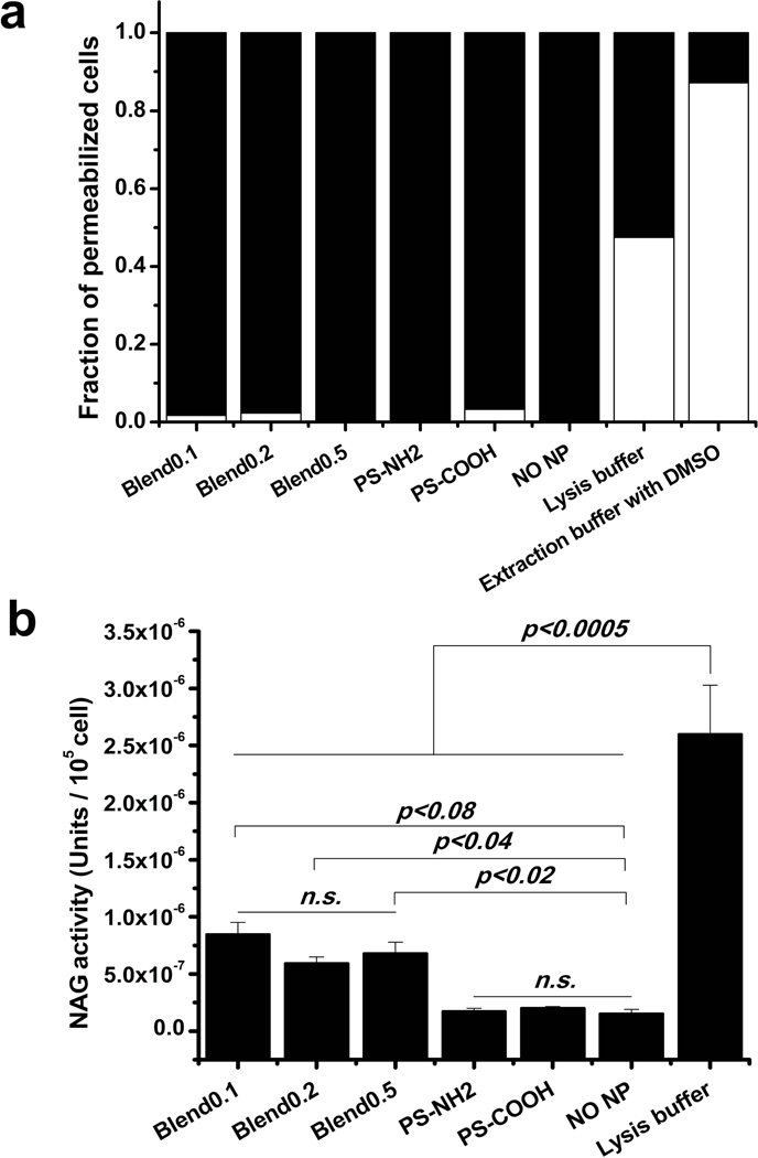
Fig 5. The release of lysosomal enzyme, N-acetyl-β-D-glucosaminidase (NAG), mediated by blend particles.
DC2.4 cells were exposed to blend particles or PS particles for 4 h at 37°C, and then treated with digitonin. The fraction of permeabilized cells assessed by trypanblue staining for each cell types (blend0.1, blend0.2, blend0.5, PS-NH2, PS-COOH) (a). Black: permeabilized cells; white: membrane-intact cells. Cytosol NAG activity was measured with β-N-Acetylglucosaminidase assay kit and expressed as enzyme units per 105 permeabilized cells (b).

