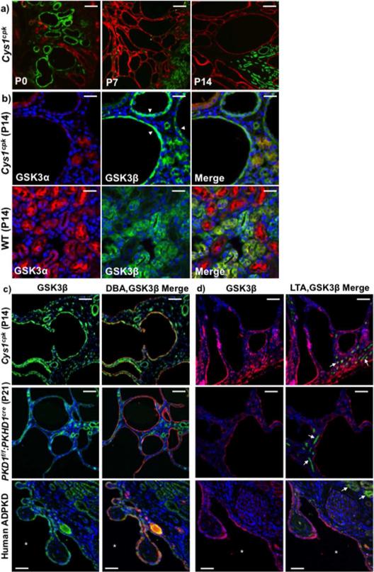
Fig 2. GSK3β is expressed in cyst lining epithelium.
(a) Cys1cpk kidneys show LTA (green) staining cysts at P0 and DBA (red) staining cysts at P7 and P14. Scale bar=50μm. (b) At P14, Cys1cpk kidneys show GSK3β (green) staining in cyst-lining epithelium (white arrow heads) and GSK3α (red) in non-cystic tubules. In wild type mice show ubiquitous staining for GSK3α and GSK3β in renal tubules. Scale bar=25μm (c) Immunofluorescence staining for GSK3β (green) and DBA (red, representing collecting ducts) and (d) GSK3β (red) and LTA (green, representing proximal tubules-white arrows) in Cys1cpk, PKD1f/f:PKHD1cre mice and human PKD kidneys. DAPI (blue) staining indicates nucleus in all figures. Scale bar=50μm.

