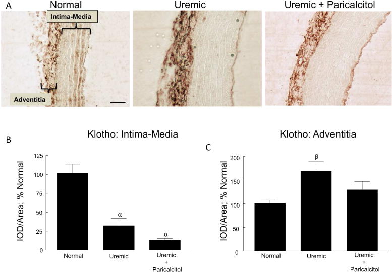
Figure 3.

Vascular Klotho. (A) Representative images of Klotho immunostaining of normal, uremic, and uremic paricalcitol-treated rat aorta, 400x; bar represents 50 microns and applies to all figures. The areas of the intima-media and adventitia are indicated in the first panel. (B) Quantitation of immunostaining of the intimal-medial area. (C) Quantitation of immunostaining of the adventitia. αp≤0.001 versus Normal, βp≤0.05 versus Normal; n=6 each; Average ± s.e.m.
