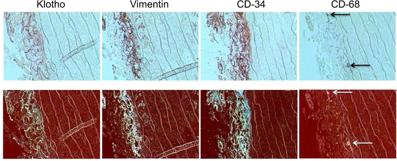
Figure 6.

Localization of Klotho, Fibroblasts, Undifferentiated Fibroblasts and Macrophages in Uremic Rat Aorta. (Upper panels) Representative images of staining for Klotho, fibroblasts (vimentin), undifferentiated fibroblasts (CD-34) and macrophages (CD-68) in uremic rat aorta; 400x. (Lower panels) Images were contrast-enhanced and inverted to better visualize staining (seen in white).
