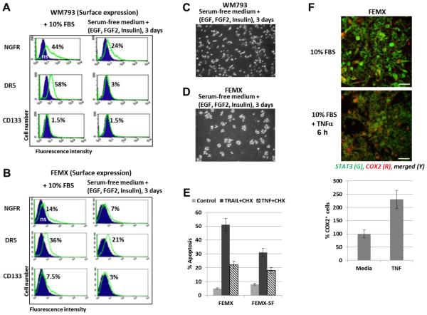
Fig. 7. Surface and total expression of receptors and TRAIL-induced apoptosis in WM793 and FEMX melanoma cells that were grown as adherent (media with 10% FBS) or suspension (serum-free media with supplements) cell cultures.
(A and B) Surface expression of receptors in WM793 and FEMX cells after culture in the complete media or growth in serum-free media with supplements: EGF (20 ng/ml, FGF2 (20 ng/ml) and insulin (5 μg/ml). Immunostaining receptors and the flow cytometry were performed. (C and D) Spheroid culture of FEMX cells and mixed (adherent and floating) culture of WM793 were observed after culturing in serum-free media with supplements. (E) TRAIL+CHX and TRAIL+TNF-induced apoptosis was induced by 24h-treatment of FEMX cells grown before treatment in the complete media (for 2 days) or serum-free (SF) media with supplements (for 3 days). PI staining of the cell nuclei and FACS analysis were performed for the detection of apoptotic levels. (F) Effects of TNFα (20 ng/ml) on expression levels of COX2 8 h after treatment of FEMX cells. Confocal images are shown after immunostaining with anti-STAT3 (green) and anti-COX2 (red) Abs. The bottom panel demonstrates relative increase of COX2-positive cells after TNF treatment.

