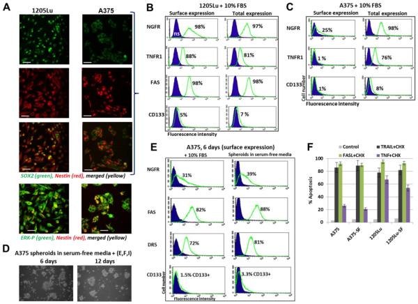
Fig. 8. Metastatic melanoma lines, 1205Lu and A375, express stem cell and proapoptotic markers.
(A) Confocal analysis of 1205Lu and A375 metastatic cells after immunostaining using rabbit polyclonal Ab to SOX2, a pluriopotency marker (green) and monoclonal antibody to Nestin , an early neuroprogenitor marker (red). Bar = 50 μm. An additional immunostaining with anti-ERK-P and anti-Nestin Abs was also performed. (B and C) Surface and total expression levels of NGFR, TNFR1, CD133, FAS and DR5 in A375 and 1205Lu melanoma cells. Percentage of positive cells is indicated. Total expression levels in 1205Lu and A375 cells were determined after permeabilization of the plasma membranes with 0.5% NP40 and immunostaining followed by FACS analysis. (D and E) A375 spheroids in serum-free media with supplements: EGF, FGF2 and insulin. Surface expression of receptors in A375 spheroids is shown on panel E. (F) Comparison of apoptotic levels in A375 cell and 1205Lu cells after culturing in the complete or serum-free (SF) media with supplements.

