Figure 1.
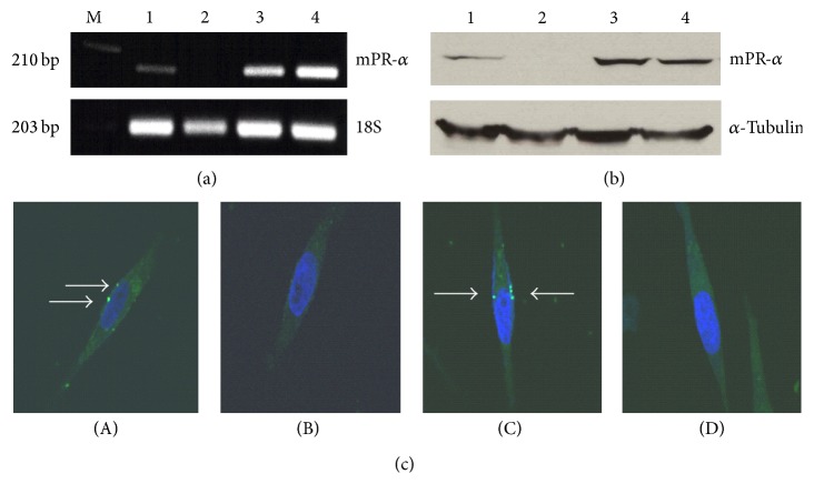
MPRα mRNA (a) and protein (b) expressions and receptor distribution (c) in various human BPBC cells. As shown in (a) and (b), the samples of mPRα mRNA and protein are indicated as “1–4,” representing MB231Br, MB231, mPRα cDNA transfected MB231 (231 w/mPRα), and MB468 breast cancer cells, respectively. The expressions of 18S and α-tubulin are used as references. (c)(A)–(c)(C) show the results of incubating the cells with P4-BSA-FITC conjugates (white arrows indicate the specific binding sites), MB231Br cells, MB231 cells, and MB231 cells w/mPRα, respectively; (c)(D) shows the result of incubating MB231Br cells with P4-BSA-FITC conjugates and excessive free P4. Images were taken under confocal microscope using ×60 oil objective lens.

