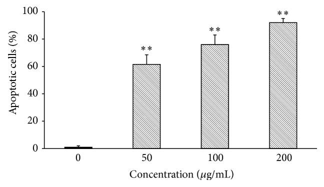
Figure 5.

Flow cytometric analysis of apoptosis in MRE-treated HepG2 cells. HepG2 cells were incubated for 24 h with 50, 100, and 200 μg/mL of methanolic root extract and apoptosis was assessed by Annexin V/7-AAD double staining. Values are means ± SD of three replicates from three independent experiments. Significant differences between the control versus treated cells are indicated by ∗∗(P < 0.01).
