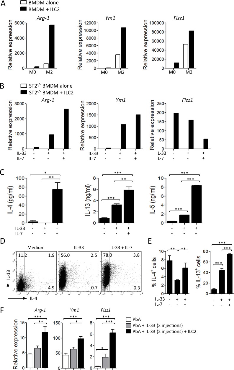
Figure 5. ILC2 promote M2 macrophage polarization.
(A) BMDM from C57BL/6 mice were cultured in the lower chamber of a 24-transwell plate in complete medium alone (M0) or supplemented with IL-4 (M2). In some experiments ILC2, sorted from naïve WT mice pre-treated with IL-33, were added to the upper chamber. After 48 h, BMDM were collected and assayed for the expression of M2 markers by qPCR (relative to Hprt1). (B) ST2-deficient BMDM were co-cultured in transwell plates as above with WT ILC2 in the presence of IL-33 alone or in combination with IL-7. After 48 h, BMDM were collected and assayed for the expression of M2 markers by qPCR (relative to Hprt1). Type 2 cytokines in the supernatants of ILC2 cultured in the presence of IL-33 or IL-33 + IL-7 were determined by ELISA (C), or by FACS (D, E). Data are mean ± SEM (n = 3 per group), representative of two independent experiments, *P<0.05, **P<0.01, ***P<0.001 by two-tailed ANOVA. (F) ILC2 sorted from mice pre-treated with IL-33 were adoptively transferred (2×10⁶ cells, i.v., on day −1) into naïve C57BL/6 mice which were infected with PbA (104 pRBCs, i.v., on day 0). The recipients, were given 2 injections of IL-33 (0.2 μg, i.p.) 30 min and 24 h after cell transfer. Expression of Arg-1, Ym1 and Fizz1 mRNA in the spleen was measured by qPCR (relative to Hprt1) on day 7. Data are mean ± SEM (n = 5 per group) *P<0.05, **P<0.01, ***P<0.001 by two-tailed ANOVA.

