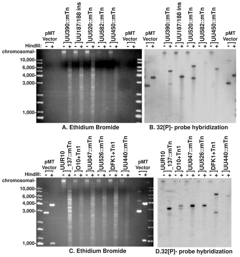
Figure 2.
In gel hybridization detection of gentamicin selection gene. Agarose gel separation of total genomic DNA extracted from transposon mutated Ureaplasma strains. Comparing HindIII digested and undigested genomic DNA. A and C show ethidium bromide visualisation of the DNA prior to probing with the gentamicin resistance gene (visualised by autoradiography in B and D). Three mutants (UU350::mTn, DFK1+Tn1, and O10+Tn1) show 2 bands suggesting a mixed colony or 2 insertion sites. No undigested samples show any extra-chromosomal plasmid DNA. All the remaining examined isolates show a single insertion site into the genome. HindIII-digested and undigested pMT85 vector is shown along with the KAPA Universal DNA ladder for fragment size comparison.

