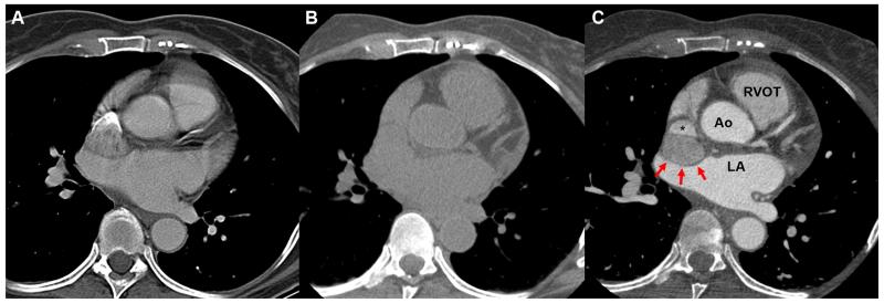
Figure 1.
Different scanning methods with CT. (A) A contrast chest CT that is not gated to the cardiac cycle demonstrates the blurriness of the cardiac silhouette. Contrast can be seen entering into the superior vena cava. (B) Non-contrast gated CT demonstrates the difficulty in seeing the mass next to the superior vena cava. (C) A contrasted gated CT allows better visualization of the mass (red arrows) impinging onto the superior vena cava without blurring of the cardiac silhouette. Ao: Aortic root, LA: left atrium, RVOT: right ventricular outflow tract.

