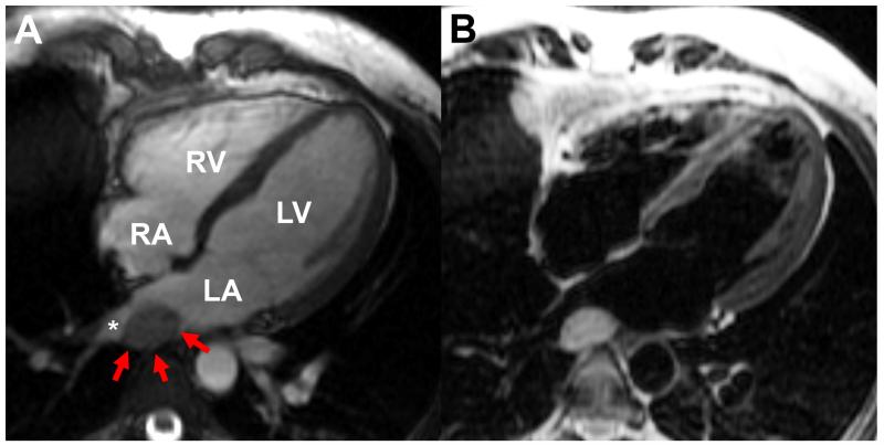
Figure 2.
(A) Cardiac MRI steady state free precession image in the 4-chamber view demonstrating an oval mass (red arrows) posterior to the left atrium (LA) abutting the right lower pulmonary vein (asterisk); (B) with turbo spin echo, the blood signal can be nulled to enhance the appearance of surrounding structures such as the mass (B). LA: left atrium, LV: left ventricle, RA: right atrium, RV: right ventricle.

