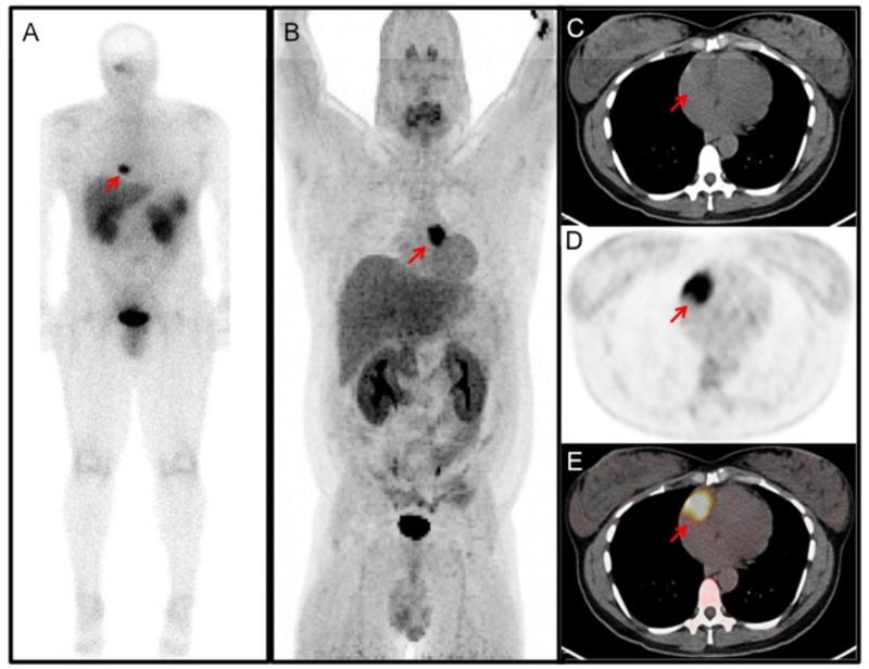
Figure 3.
Functional imaging of cardiac PGLs from different patients. (A) Octreotide (111In-pentetreotide) scan demonstrating increased uptake in the neck and heart. There is also increased uptake in the liver, kidneys, spleen, gallbladder, and urinary bladder, which are physiologic. (B) 18F-FDOPA (18F-fluorodihdyroxyphenylalanine) PET demonstrating a large cardiac paraganglioma at the base of the heart near the root of the great vessels. (C-E) 18F-FDG PET/CT of a mass in the right atrioventricular groove. (C) Non-contrast CT scan for localization; (D) 18F-FDG PET showing an area of high activity; (E) fused PET/CT image.

