
Fig. 2.
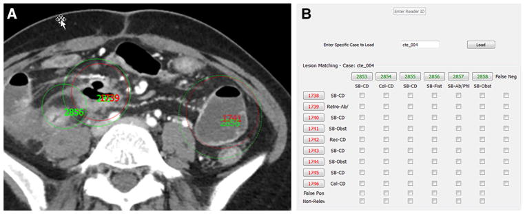
Image showing matching of reader (green circles and numbers) and reference (red circles and numbers) marking viewed by two GI radiologists not participating in CTE interpretation. Radiologists could scroll up and down through the dataset to determine if inflamed segments could were marked by reference physicians and radiologists readers (A). Each reference and reader marking was given a unique reference identification number. A table (B) then permitted matching of reader and reference markings. Unmatched reference identification numbers were labeled as false negatives, and unmatched reader markings were labeled as false positive or non-relevant (e.g., if marking a renal mass rather than a bowel finding).