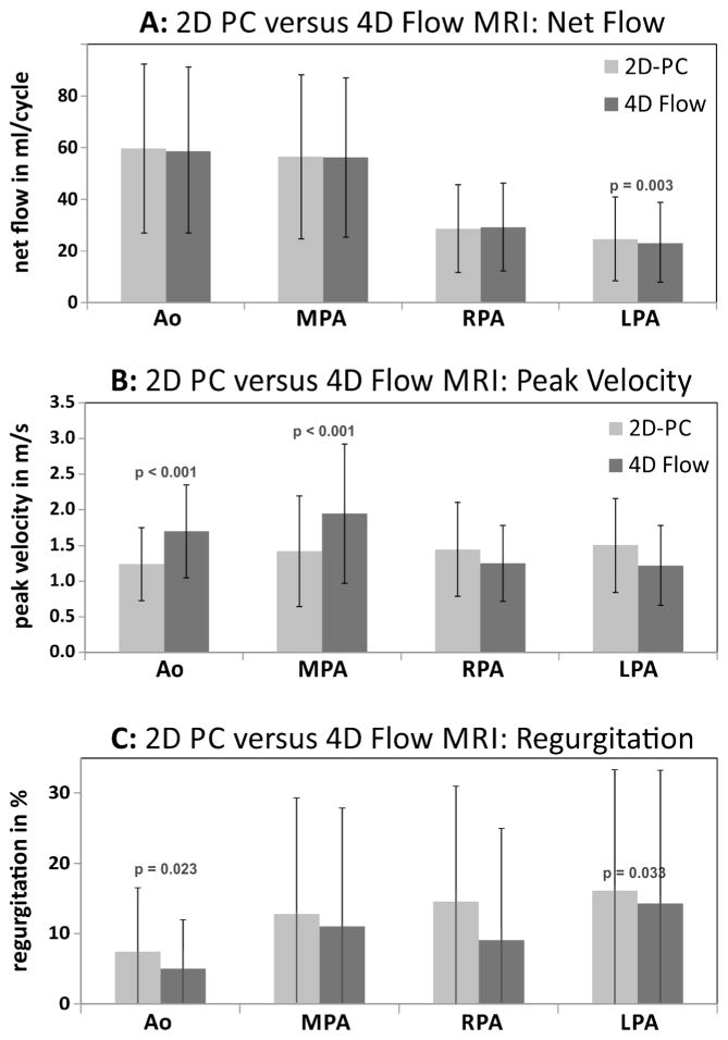
Fig. 2.
Graphs show comparison of aortic and pulmonary flow parameters between 2-D phase-contrast (PC) MRI and quantification based on 4-D flow. Comparisons are for (a) net flow, (b) peak systolic velocity and (c) regurgitant fraction. Volumetric flow analysis (Fig. 1) in the aortic root/ascending aorta was used for 4-D flow-based peak systolic velocity quantification (b) in the aortic root/ascending aorta and MPA. Ao aorta, LPA left pulmonary artery, MPA main pulmonary artery, RPA right pulmonary artery

