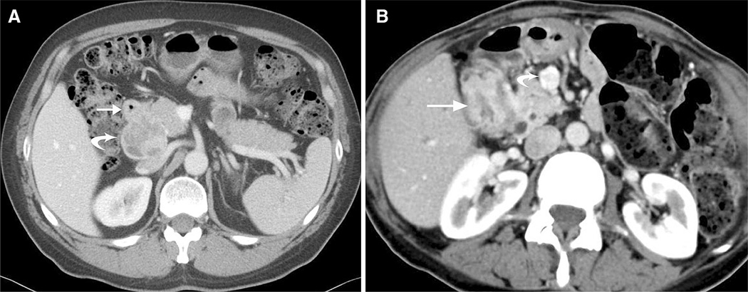
Fig. 2.
A 51-year-old man with incidental neuroendocrine tumor discovered following trauma. Axial CT image of the upper abdomen in the venous phase shows wall thickening of the first portion of duodenum (arrow) without obvious focal mass and a 5.0 × 4.8 cm enhancing and partially necrotic portal caval lymph node (curved arrow). Final pathology revealed a 1.7-cm neuroendocrine tumor of the duodenum and metastatic lymphadenopathy. B 60-year-old female with neurofibromatosis type 1. Axial CT images demonstrate a longer 3.3-cm segment of wall thickening (arrow pathologically proven well-differentiated neuroendocrine tumor with features of a somatostatinoma) and enhancing lymphadenopathy in the root of the mesentery (curved arrow).

