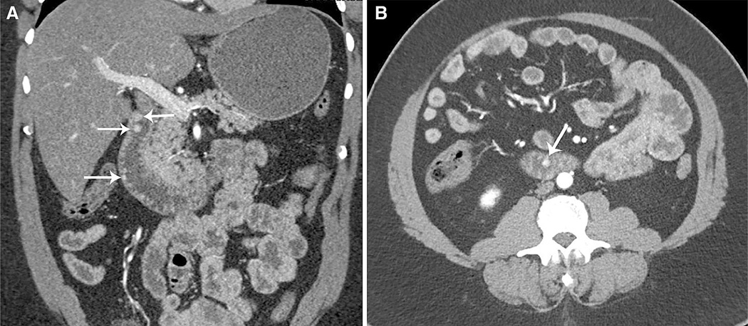
Fig. 4.
57-year-old man with nonspecific reflux and mild anemia and multifocal carcinoid tumors. Coronal (A) and axial (B) CT images in the arterial phase show multiple tiny duodenal well-differentiated carcinoid tumors (arrows). Patient did not have a history of clinical syndrome such as MEN-1 or NF-1.

