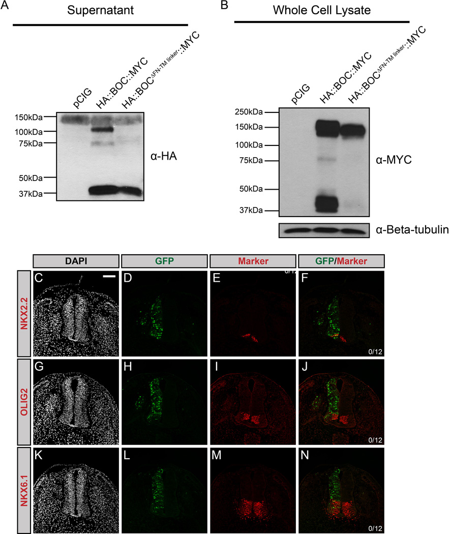
Figure 3. BOC undergoes extracellular proteolytic cleavage mediated by a juxtamembrane domain.
(A) Western blot analysis of supernatants from COS-7 cells transfected with pCIG, HA::Boc::MYC, or HA::BocΔFN-TM linker::MYC, probed with anti-HA antibody. Note the presence of a 100kDa band in the HA::BOC::MYC lane, but not the HA::BOCΔFN-TM linker::MYC lane, while a 37kDa band is detected in both lanes. (B) Western blot analysis of whole cell lysates probed with anti-MYC antibody and anti-Beta-tubulin as a loading control. (C–N) Forelimb level transverse sections of Hamburger-Hamilton stage 21–22 chicken neural tubes electroporated with BocΔFN-TM linker, and stained with DAPI (grayscale; C, G, K), and antibodies against NKX2.2 (red; E, F), OLIG2 (red; I, J), or NKX6.1 (red; M, N). GFP+ cells denote electroporated cells (green; D, H, L). Merged images are shown at the right, including quantitation of the number of embryos that display ectopic NKX2.2, OLIG2 or NKX6.1 expression. Scale bar (C), 10µm.

