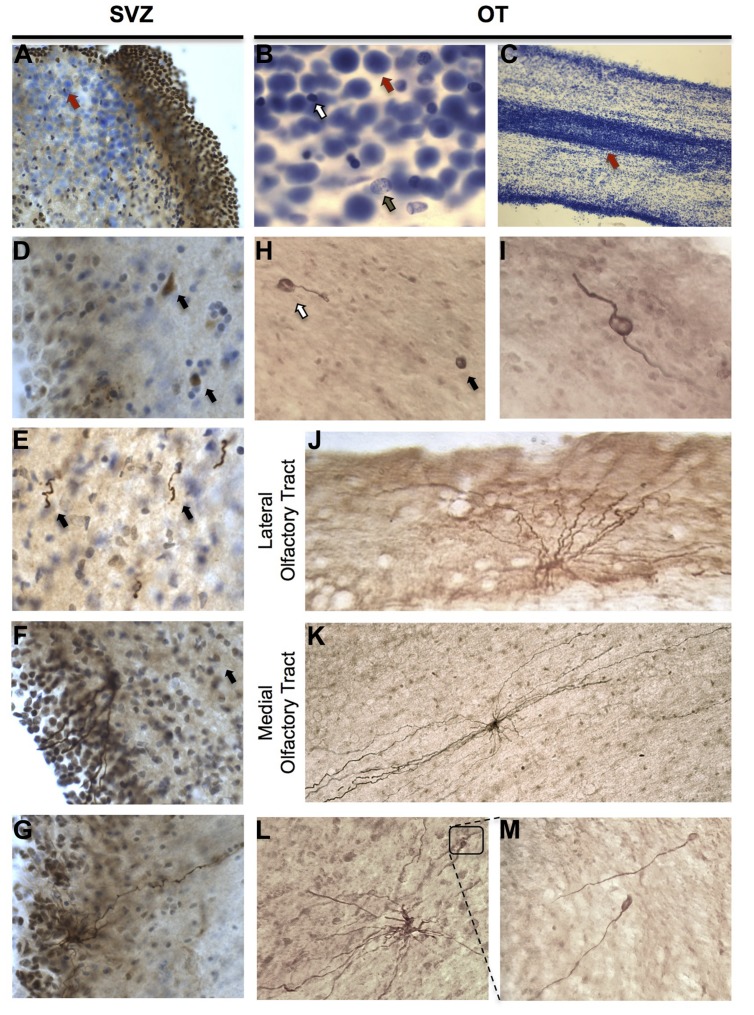
FIGURE 2.
Cellular composition and characterization of DCX-immunoreactive (IR) cells in the human olfactory tract (OT) and SVZ. (A–C) Large, densely packed cells of unknown identity were identifiable in SVZ and OT regions and formed a corridor suggestive of a rostral migratory stream. When stained with cresyl violet, these cells (red arrows) were readily distinguishable from both glia (white arrow) and neurons (brown arrow). (D–G) DCX-IR expressing cells in the human SVZ displayed variable morphologies including: neuroblast-like cells lacking processes (arrows in D); DCX-IR cells with leading and/or trailing processes possessing a rostrocaudal orientation (arrows in E); and multipolar cells possessing multiple processes that extended toward the ventricular surface and, occasionally, a additional basal process which projected in the opposite direction. (H,I) In the OT, DCX-IR somata with (white arrow) and without (black arrow) processes were observed, as were DCX-IR bipolar cells. (J,K) DCX-IR multipolar cells were present in both lateral and medial regions. DCX-IR processes were generally aligned parallel to the tract, particularly in medial OT regions. (L,M) Occasionally, DCX-IR multipolar cells possessed bulbous, growth cone-like tips.

