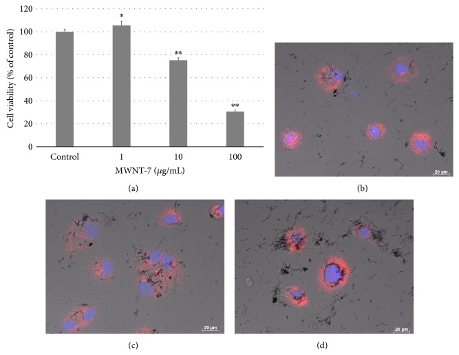
Figure 2.
HMCs were exposed to MWNT-7. (a) Viability of HMCs exposed to variety concentration of MWNT-7 in 0.1% gelatin for 24 h. P values were compared to HMCs exposed to MWNT-7 in dispersant control. Mean ± S.D. n = 6, ∗ P < 0.05, ∗∗ P < 0.001. Image of HMCs exposed to 10 μg/mL MWNT-7 in 0.1% gelatin at 2 h (b), 6 h (c), and 24 h (d). DIC and fluorescence images were merged. Nuclei were stained blue with H33342 and lysosome were stained red with CytoPainter.

