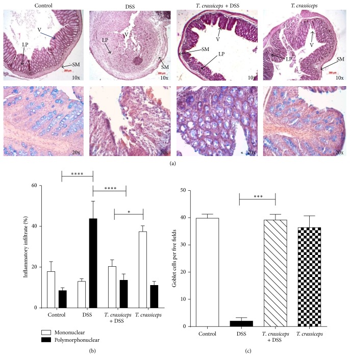
Figure 2.
T. crassiceps-infected mice do not display severe pathology during ulcerative colitis. (a) Upper panel, colon tissue histology stained with H&E and showing colonic inflammation in different groups: magnification is 10x; bottom panel, Alcian blue-stained goblet cells (blue): magnification is 20x. (b) Percentages of neutrophils and monocytes located in distal colons. (c) Number of goblet cells; these cells were quantified from at least 20 crypts per region in five fields in four different slides per animal. Data are means ± SEM. * P < 0.05, ** P < 0.01.

