
Figure 2.
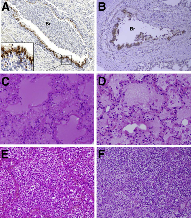
Representative pathological changes during fatal primary influenza and secondary bacterial infections in human autopsies from the 1918 (A, C, and E) and 2009 (B, D, and F) pandemics. A: Immunohistochemical stained section of lung from a 1918 pandemic influenza fatal case showing acute influenza viral bronchiolitis with infiltration of neutrophils and other inflammatory cells in the lumen of a bronchiole (Br). Influenza viral antigen (reddish-brown stain) is readily apparent in the apical cells of the bronchiolar respiratory epithelium (inset), on a hematoxylin-stained background.50B: Immunohistochemical stained section of lung from a 2009 pandemic influenza fatal case showing acute influenza viral bronchiolitis (Br). Influenza viral antigen (reddish-brown stain) is readily apparent in the apical cells of the bronchiolar respiratory epithelium, on a hematoxylin-stained background.51C: Hematoxylin and eosin (H&E)–stained section of lung from a 1918 pandemic influenza fatal case showing diffuse alveolar damage with hyaline membranes lining alveoli. The alveolar airspaces contain edema fluid, strands of fibrin, desquamated epithelial cells, and inflammatory cells.50D: H&E-stained section of lung from a 2009 pandemic influenza fatal case showing diffuse alveolar damage with hyaline membranes lining alveoli. The alveolar air spaces contain edema fluid, strands of fibrin, desquamated epithelial cells, and inflammatory cells.51E: H&E-stained section of lung from a 1918 pandemic influenza fatal case showing a massive infiltrate of neutrophils that fills the alveolar air spaces associated with a secondary bacterial bronchopneumonia. Alveolar capillary congestion is prominent.6F: H&E-stained section of lung from a 2009 pandemic influenza fatal case showing a massive infiltration of neutrophils in the airspaces of alveoli associated with a secondary bacterial bronchopneumonia.51A and C were modified from Sheng et al (published by College of American Pathologists),50E from Taubenberger and Morens (published by Annual Reviews),6 and B, D, and F from Gill et al (published by National Academy of Sciences of the United States of America).51 All images used with permission of the publishers. Original magnifications: ×40 (A and B); ×100 (E and F); ×200 (C and D).