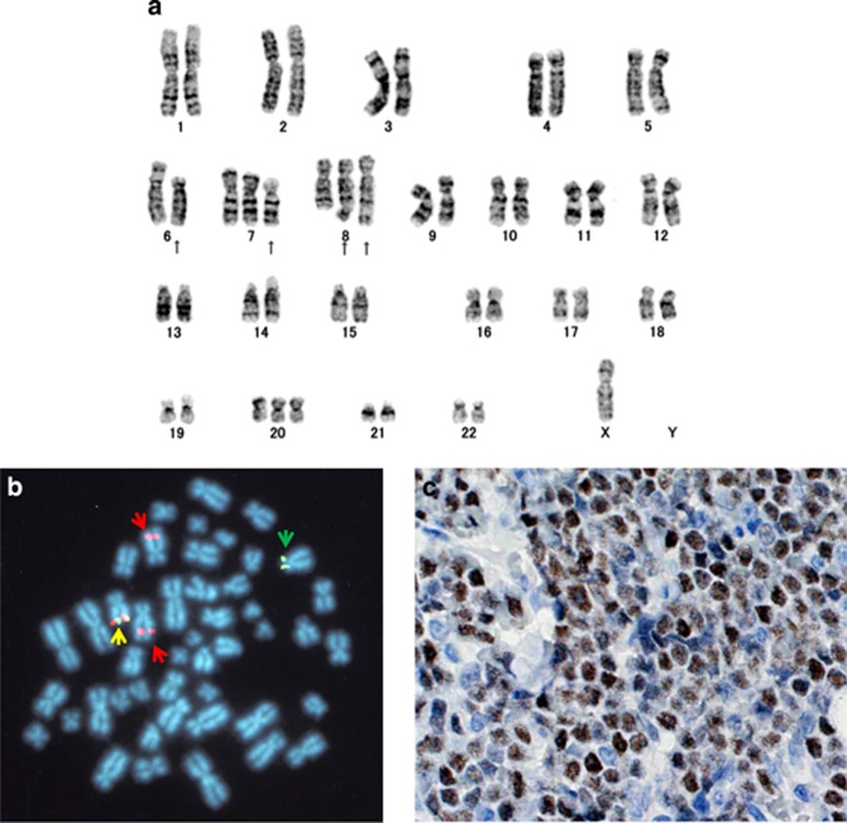
Figure 1.
(a) G-banded karyogram of the patient's BM cells. Arrows indicate the aberrant chromosomes. (b) Fluorescence in situ hybridization analysis of BM cells using Spectrum Orange-labeled 5' LSI MYC and Spectrum Green-labeled 3' LSI MYC probes. Normal chromosome 8 is shown by fusion of the two probes (a yellow arrow). Derivative 6 and 8 chromosomes are indicated by green and red arrows. (c) Immunohistochemistry of biopsied LN using anti-MYC antibody. MYC was highly expressed in nucleus of tumor cells.

