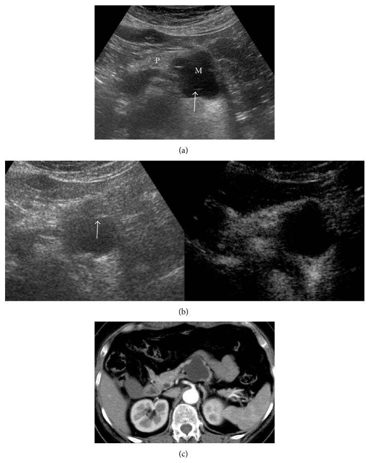
Figure 2.
Pancreatic lesion was found by physical examination and was diagnosed as MCN by surgical pathology. (a) Cystic lesions in the tail of the pancreas were indicated by US (M: mass; P: pancreas), with multiple septa (arrow). The case was diagnosed as type III. (b) Enhancement was not shown in cystic lesions in CEUS (the right picture). The case was diagnosed as type I. (c) Enhanced CT indicated no enhancement in the cystic lesion. The case was diagnosed as type I.

