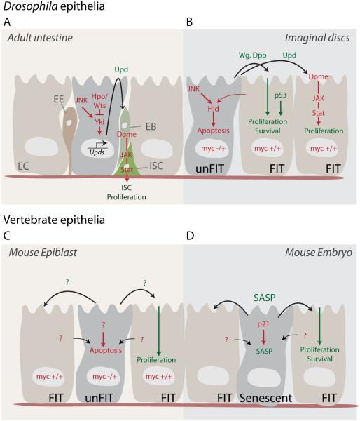
Figure 1. Paracrine signaling in the control of tissue regeneration, homeostasis and remodeling.
(A) In the posterior midgut epithelium of Drosophila, damaged enterocytes (EC) secrete Upd cytokines upon the activation of stress signaling involving JNK and Yki. Upds activate the JAK/STAT signaling pathway of intestinal stem cells (ISCs), inducing their proliferation and generating enteroblasts (EB), which go on to differentiate into ECs or enteroendocrine (EE) cells.
(B) In imaginal discs, cell competition results in the induction of apoptosis in slow growing cells (‘unfit cells’, here exemplified by cells with a lower dose of myc). Faster growing (‘fit’) cells induce apoptosis in unfit cells in a p53 and JNK-dependent manner. Apoptotic cells, in turn, promote compensatory proliferation by inducing Dpp, Wg and Upds.
(C) In the mouse epiblast, fit MYC over-expressing cells induce apoptosis and engulfment of unfit cells, which in turn promote the proliferation of fit cells through paracrine signals.
(D) In the mouse embryo, activation of the cell cycle inhibitor p21 can induce senescence. Senescent cells, through the SASP, fine-tune patterning and growth by supporting the survival and proliferation of neighboring fit cells.

