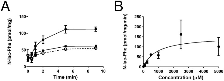
Fig. 3.
N-lac-Phe is transported into inside-out membrane vesicles by ABCC5. (A) Control vesicles (▪) and ABCC5-containing vesicles (○ and ●) were incubated with 100 μM N-lac-Phe at 37 °C in the presence (solid line) and absence (dashed line) of 5 mM ATP. At the indicated time points, a sample containing 75 μg of protein was taken. After washing over a filter, the vesicular content was analyzed by LC/MS (n = 3–6). (B) Concentration dependence was assessed by incubating control and ABCC5-containing vesicles with several concentrations of N-lac-Phe in the presence of ATP and determining ABCC5-dependent uptake after 2 min (n = 3–4 for concentration <1,000 μM and n = 8 for concentration ≥1,000 μM). The data were fitted to Michaelis–Menten kinetics (solid line) using GraphPad Prism. Data are presented as mean ± SEM.

