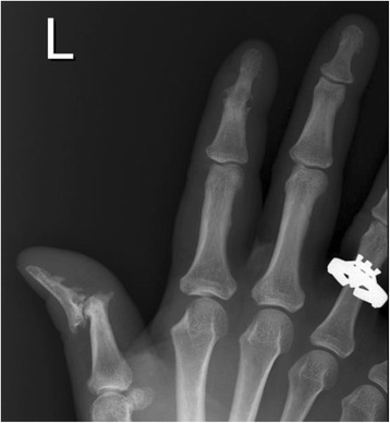
Fig. 2.

X-ray image of changes in bones observed in psoriatic arthritis. Bone changes in psoriatic arthritis (PsA) patients may differ between patients and may also differ within the same patient. The heterogeneity observed within a PsA patient is illustrated. Left-hand radiograph from a PsA patient showing severe erosive disease and subluxation at the first distal interphalangeal (DIP) with fluffy periosteal new bone formation on the terminal phalange. Ankylosis of the second DIP joint is also demonstrated
