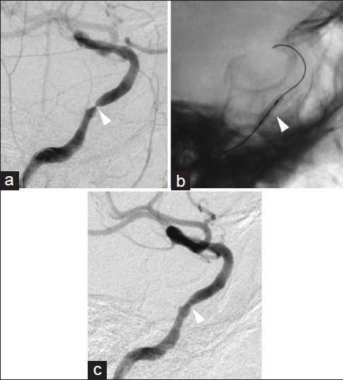
Figure 4.

Pretreatment and posttreatment right ICA angiograms. The pretreatment angiogram indicating severe stenosis in the cavernous segment of the ICA (a, arrowhead). A microguidewire was advanced through the stenotic lesion and into the supraclinoid segment of the ICA. Inflation of a contrast-filled gateway PTA balloon (b, arrowhead) within the stenotic cavernous segment of the ICA dilated the vessel lumen, improving flow (c, arrowhead)
