Abstract
Introduction:
The objective of this study was to evaluate dentinal defects formed by new rotary system — Protaper next™ (PTN).
Materials and Methods:
Sixty single-rooted premolars were selected. All specimens were decoronated and divided into four groups, each group having 15 specimens. Group I specimens were prepared by Hand K-files (Mani), Group II with ProTaper Universal (PT; Dentsply Maillefer), Group III with Hero Shaper (HS; Micro-Mega, Besancon, France), and Group IV with PTN (Dentsply Maillefer). Roots of each specimen were sectioned at 3, 6, and 9mm from the apex and were then viewed under a stereomicroscope to evaluate presence or absence of dentinal defects.
Results:
In roots prepared with hand files (HFs) showed lowest percentage of dentinal defects (6.7%); whereas in roots prepared with PT, HS, and PTN it was 40, 66.7, and 26.7%, respectively. There was significant difference between the HS group and the PTN group (P < 0.05).
Conclusion:
All rotary files induced defects in root dentin, whereas the hand instruments induced minimal defects.
Keywords: Dentinal defects, hand files, NiTi instruments, ProTaper next™ rotary system, root canal preparations
INTRODUCTION
The irrevocable aim of endodontics is a three-dimensional unblemished seal of the root canal system which is achieved by perfect designing of the canal diameter and canal form. The biomechanical preparation is one of the major steps for removal of bacteria and debris in the root canal so as to achieve a successful endodontic treatment.[1,2] During root canal instrumentation there are complications such as perforations, ledge formation, transportation of canal, and formation of cracks in the root dentin.[3,4] At times, in the zeal of biomechanical preparation of the canal we inevitably end up damaging the root dentin, which becomes a gateway to dentinal cracks and minute intricate fractures; thereby, causing failure of treatment.[1,5,6,7]
As a result of craze lines or microcracks, there might be occurrence of root fracture that propagates due to repeated application of stress by the occlusal forces.[7,8,11] Shemesh et al.,[12] observed more dentinal defects in teeth which were obturated with spreader than teeth obturated without spreader. In different degrees, dentinal damage can occur due to procedures like biomechanical preparation, obturation, and retreatment.[9,10,11]
Complexities in the preparation of root canal may be attributed to variation in the design of the cutting instrument, taper, or composition of the material from which it is made.[2] Hand instrumentation — the milestone of endodontic practice in the past — though have lost popularity, still remain the integral part of canal preparation.[3]
Rotary instrument by its innate behavior in the canal may result in more friction, which may increase dentinal defects and microcracks formation in comparison to hand instruments.[13,14] Possible relationship between the design of NiTi rotary instruments and the incidence of the vertical root fractures was found by Kim et al.,[14] and it was concluded that the design of the file affects strain concentration and the apical stress during instrumentation of root canal.
Recently, the ProTaper next (PTN, Dentsply, Maillefer) files were introduced in the family of NiTi rotary instruments with a completely new design comprising of unique swaggering movement, greater flexibility, the M-wire technology, the 5th generation of continuous improvement, and its offset design.[9,10,15]
Whether it is rotary or hand files (HFs), they are assumed to cause limited frictional forces within the canal, hence creating dentinal defects. So there is need to study the behavior of different NiTi rotary instruments and the newly developed rotary system, PTN, on root dentin.
MATERIALS AND METHODS
Sixty single-rooted premolars were selected and stored in purified water. Teeth with curved roots, calcified canals, extracanals, and teeth with developmental anomaly or resorption were excluded from the study. The teeth were decoronated at coronal portions by using a diamond disc, leaving roots approximately of 10mm in length. All the roots were inspected with transmitted light for detecting any preexisting cracks or any craze-lines by using a stereomicroscope under ×12, to exclude teeth with such findings from this study.
Patency of the canal was established using a #10 K-File (Mani, Japan) in the canal. The specimens were then divided into four groups; each group containing 15 specimens each.
Group I: HFs.
Group II: Protaper Universal (PT).
Group III: Hero Shaper (HS).
Group IV: PTN.
In the HF group, HFs upto file #40 were used for canal preparation. In the PT (Dentsply, Maillefer), HS (Micro-mega, Besancon, France), and PTN (Dentsply, Maillefer) groups; preparation of the canals was done using speed and torque controlled motor (X-SMART; Dentsply, Maillefer).
In the Hand Files Group, step-back technique was used upto file #40. In the PT group, the following sequence of PT rotary NiTi files were used for preparation of canals at 300 rpm: The shaping file X for coronal enlargement, and S1, S2, F1, F2, and F3 files, corresponding to apical size 30, used at the working length. In the HS group, the HS NiTi files were used upto file #30 at 300 rpm in crown-down sequence. In PTN group, the PTN rotary system files were used at 300 rpm in the following sequence: X1, X2 and X3, corresponding to apical size #30. The PTN rotary files were used in a constant rotation at a speed of 300 rpm with light apical pressure (recommended torque is 2.0 Ncm, adjustable up to 5.2 Ncm according to practitioner experience).
Flutes of the instruments were cleaned frequently to check any signs of distortion or wear. The PTN instruments are recommended to be used mechanically (manually in very severe curvatures) in a clockwise continuous motion with a brushing motion, away from external root concavities, to facilitate flute unloading and apical file progression.[16,18]
In all the experimental groups, irrigation of each specimen was done with sodium hypochlorite solution.[11]
Sectioning and Microscopic Evaluation
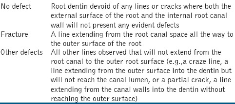
Sectioning of all the roots was done perpendicular to the long axis at 9, 6, and 3 mm using a diamond disc under water cooling.[5,7,11] Digital images of each sectioned root was captured using a ×40 stereomicroscope by using a digital camera (Olympus, Tokyo, Japan). Two operators checked each specimen for the presence of dentinal defects. Roots were classified as “no defect”, “fracture”, and “other defects” as described in Table 1.
Table 1.
Classification for identification of defects in the specimens

Statistical Analysis
The results were expressed as the number and percentage of defects in each group. Chi-square test was used for the statistical analysis of the groups. The level of significance was set at P = 0.05 using Statistical Package for Social Sciences (SPSS) 20.0.
RESULTS
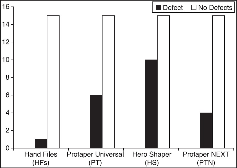
Figure 1 is a bar chart representing the number of root defects in each group. HFs group showed lowest defect (1/15) followed by PT (6/15), HS (10/15), and PTN (4/15). Statistical significant difference was seen between HFs and HS group and between HS and PTN groups (P < 0.05). No significant difference was found between the PT and HS (P > 0.05).
Figure 1.

Bar chart representing number of root defects in each group
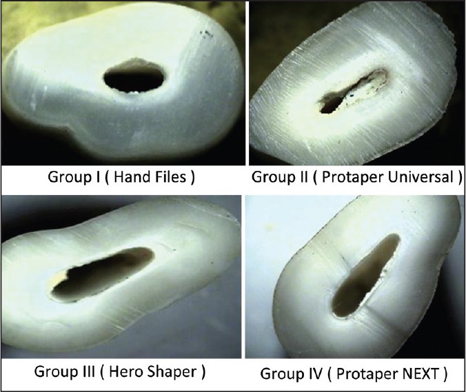
The Stereomicroscopic images of Group I, II, III and IV are shown in Figure 2.
Figure 2.

Stereomicroscopic images showing dentinal defects seen in Groups I, II, III, and IV, showing craze lines seen in Group I. Fracture and other defects in Group II craze lines and partial crack in Group III and craze lines in Group IV
DISCUSSION
In the present study; in HFs, PT, HS, and PTN, the number and incidence of defects observed in the root dentin was found to be 1/15 (6.67%), 6/15 (40%), 10/15 (66.67%), and 4/15 (26.7%), respectively. Group I (HFs) showed the lowest incidence of defects (6.67%); whereas, HS group showed the maximum incidence of defects (66.67%) as compared to other groups. The results of our study are in accordance with Imam,[7] who reported lowest number of defects (1/20) by HFs; and Yold as et al.,[11] observed highest incidence of defects (12/20) by HSs rotary files. Similar to results by Capar et al.,[19] in the present study the number and percentage of defects shown by the PTN rotary files were 4/15, that is, 26.7%. The results of the present study are not in accordance with the results by Bier et al.,[5] where the HFs showed no defect and PT rotary files showed highest incidence of dentinal damage (16%).
Excess removal of root dentin during root canal preparation and obturation of the canal with spreader may create fracture in the teeth. The important goal in endodontics is resistance to tooth fracture because such fractures might cause decrease in the long-term survival rate.[11,12] In the presents study, only one dentinal defect was seen in the HF group. The number of rotations required for complete root canal preparation is more with NiTi rotary instruments than with the HFs.[7,17]
Kim et al.,[14] stated that taper of the files is the responsible for increase of stress on the walls of the root canal; whereas, Bier et al.,[5] stated taper of the files as one of the contributing factor for crack formation in root dentin. PT have more taper (0.07, 0.08, and 0.09, respectively) than the HSs (0.04 and 0.06) and the PTN (X1, X2, and X3; 0.04, 0.06, and 0.07, respectively). This explains that there can be formation of cracks in the PT group, as reported earlier by Bier et al.,[5] Liu et al.,[20] Barreto et al.,[21] and Liu et al.[22] Furthermore, relatively low flexibility of the HS may have contributed to the maximum number of defects in HS group in the present study.[7]
Rotational force is applied to the canals of the root by NiTi rotary instruments, thus creating craze line or microcracks in root dentin. Formation of such defects may be associated with the design of tip, cross-sectional geometry, taper type (constant or progressive), flute form, and pitch (constant or variable).[7,12]
The PT files have a triangular cross-sectional geometry, HS having a triple helix cross-sectional geometry; whereas, the PTN is rectangular. There is variable pitch in both PT and PTN files. Thus, it can be stated that design of the rotary files is not the only factor for defect formation in root dentin. Lam et al.,[23] stated that forces shaping the root dentin can be affected by the file design. Risk of root fracture is increased due to the forces generated during the root canal preparation.[14] PTN files have M-wire technology with off-centered rectangular cross-section, giving the file a snake-like swaggering movement as it moves along the root canal, thus reducing the screw effect, the unwanted taper lock, and torque on any of the given file; thus decreasing the file-root dentin contact.[24,25]
M-wire alloy NiTi material with controlled memory NiTi wire are flexible than those made from conventional NiTi wire.[19] Thus, such flexibility of PTN rotary files may have contributed in less number of dentinal defects formation as compared to PT and HS. Capar et al.,[19] concluded that the swaggering motion and less taper of the PTN instruments could change the root canal volume to an extent as that of the higher tapered instruments.
Limitations
Use of different speed and torque settings for each rotary system could be the limitation of our study. Increase in the rotational speed is associated with increased cutting efficiency.[19]
Simulation of periodontal ligament was not done in the present study. Capar ID et al.[19] stated that simulation of the periodontal ligament is necessary for investigating the influence of forces on formation of crack or fracture strength. Moreover, the periodontal ligament has viscoelastic property. It plays an important role in stress dissipation created by application of load to the teeth.
CONCLUSIONS
Within the limitations of this in vitro study, PTN rotary system can induce less dentinal defects than PT and HS.
ACKNOWLEDGMENTS
The authors thank Mr. Ashok Bhagat (Praj Laboratories, Pune, India) for testing of the specimens, Mr. Jaydeep Nayse (Associate Professor, Department of Community Medicine, NKPSIMS, Nagpur, India) for statistical analysis, and Mr. Nandakumar C from Dentsply (Key Account Manager, Karnataka State — India) for making Protaper next™ available for us.
Footnotes
Source of Support: Nil
Conflict of Interest: None declared.
REFERENCES
- 1.Peters OA. Current challenges and concepts in the preparation of root canal systems: A review. J Endod. 2004;30:559–67. doi: 10.1097/01.don.0000129039.59003.9d. [DOI] [PubMed] [Google Scholar]
- 2.Bergmans L, Van Cleynenbreugel J, Beullens M, Wevers M, Van Meerbeek B, Lambrechts P. Smooth flexible versus active tapered shaft design using NiTi rotary instruments. Int Endod J. 2002;35:820–8. doi: 10.1046/j.1365-2591.2002.00574.x. [DOI] [PubMed] [Google Scholar]
- 3.Blum JY, Machtou P, Ruddle C, Micallef JP. Analysis of mechanical preparations in extracted teeth using ProTaper rotary instruments: Value of the safety quotient. J Endod. 2003;29:567–75. doi: 10.1097/00004770-200309000-00007. [DOI] [PubMed] [Google Scholar]
- 4.Pasqualini D, Scotti N, Tamagnone L, Ellena F, Berutti E. Hand-operated and rotary ProTaper instruments: A comparison of working time and number of rotations simulated root canals. J Endod. 2008;34:314–7. doi: 10.1016/j.joen.2007.12.017. [DOI] [PubMed] [Google Scholar]
- 5.Bier CA, Shemesh H, Tanomaru-Filho M, Wesselink PR, Wu MK. The ability of different nickel-titanium rotary instruments to induce dentinal damage during canal preparation. J Endod. 2009;35:236–8. doi: 10.1016/j.joen.2008.10.021. [DOI] [PubMed] [Google Scholar]
- 6.Kumaran P, Sivapriya E, Indhramohan J, Gopikrishna V, Savadamoorthi KS, Pradeep Kumar AR. Dentinal defects before and after rotary root canal instrumentation with three different obturation techniques and two obturating materials. J Conserv Dent. 2013;16:522. doi: 10.4103/0972-0707.120968. [DOI] [PMC free article] [PubMed] [Google Scholar]
- 7.Al-Zaka IM. The effects of canal preparation by different Niti rotary instruments and reciprocating Wave One on the incidence of dentinal defects. M Dent J. 2012;9:137–42. [Google Scholar]
- 8.Milani AS, Froughreyhani M, Rahimi S, Jafarabadi MA, Paksefat S. The effect of root canal preparation on the development of dentin cracks. Ir Endod J. 2012;7:177–82. [PMC free article] [PubMed] [Google Scholar]
- 9.El-Rahman MA, El-Moghazy S, Abd MM, El-Azeem E, Ghada E, Mohamed Eid. Comparative study of the efficacy of two newly introduced rotary nickel titanium instruments in shaping of curved root canals. Cairo Dent J. 2008;24:447–56. [Google Scholar]
- 10.Protaper next TM flexible performance. [Last accessed on 2014 Sep 09]. Available from www.dentsplymaillefer.com .
- 11.Yoldas O, Yilmaz S, Atakan G, Kuden C, Kasan Z. Dentinal microcrack formation during root canal preparations by different niti rotary instruments and the self-adjusting file. J Endod. 2012;38:232–5. doi: 10.1016/j.joen.2011.10.011. [DOI] [PubMed] [Google Scholar]
- 12.Shemesh H, Bier CA, Wu MK, Tanomaru-Filho M, Wesselink PR. The effects of canal preparation and filling on the incidence of dentinal defects. Int Endod J. 2009;42:208–13. doi: 10.1111/j.1365-2591.2008.01502.x. [DOI] [PubMed] [Google Scholar]
- 13.Schäfer E, Lau R. Comparison of cutting efficiency and instrumentation of curved canals with nickel-titanium and stainless-steel instruments. J Endod. 1999;25:427–30. doi: 10.1016/S0099-2399(99)80272-6. [DOI] [PubMed] [Google Scholar]
- 14.Kim HC, Lee MH, Yum J, Versluis A, Lee CJ, Kim BM. Potential relationship between design of nickel-titanium rotary instruments and vertical root fracture. J Endod. 2010;36:1195–9. doi: 10.1016/j.joen.2010.02.010. [DOI] [PubMed] [Google Scholar]
- 15.Ruddle CJ, Pierre M, West JD. The shaping Movement 5th Generation Technology. Dynamics. 19:4–9. [Google Scholar]
- 16.Protaper next rotary files Performance Refined. DENTSPLY Tulsa Dental Specialties. [Last accessed on 2014 Sep 11]. Available from: www.TulsaDentalSpecialties.com .
- 17.Wilcox LR, Roskelley C, Sutton T. The relationship of root canal enlargement to finger-spreader induced vertical root fracture. J Endod. 1997;23:533–4. doi: 10.1016/S0099-2399(97)80316-0. [DOI] [PubMed] [Google Scholar]
- 18.Mahran AH, Aboel-Fotouh MM. Comparison of effects of protaper, hero shapers and gates glidden burs on cervical dentin thickness and root canal volume by using multislice computed tomography. J Endod. 2008;34:1219–22. doi: 10.1016/j.joen.2008.06.022. [DOI] [PubMed] [Google Scholar]
- 19.Capar ID, Arslan H, Akcay M, Uysal B. Effects of protaper universal, protaper next, and hyflex instruments on crack formation in dentin. J Endod. 2014;40:1482–4. doi: 10.1016/j.joen.2014.02.026. [DOI] [PubMed] [Google Scholar]
- 20.Liu R, Hou BX, Wesselink PR. The incidence of root microcracks caused by 3 different single-file systems versus the ProTaper system. J Endod. 2013;39:1054–6. doi: 10.1016/j.joen.2013.04.013. [DOI] [PubMed] [Google Scholar]
- 21.Barreto MS, Moraes Rdo A, Rosa RA. Vertical root fractures and dentin defects: Effects of root canal preparation, filling, and mechanical cycling. J Endod. 2012;38:1135–9. doi: 10.1016/j.joen.2012.05.002. [DOI] [PubMed] [Google Scholar]
- 22.Liu R, Kaiwar A, Shemesh H. Incidence of apical root cracks and apical dentinal detachments after canal preparation with hand and rotary files at different instrumentation lengths. J Endod. 2013;39:129–32. doi: 10.1016/j.joen.2012.09.019. [DOI] [PubMed] [Google Scholar]
- 23.Lam PP, Palamara JE, Messer HH. Fracture strength of tooth roots following canal preparation by hand and rotary instrumentation. J Endod. 2005;31:529–32. doi: 10.1097/01.don.0000150947.90682.a0. [DOI] [PubMed] [Google Scholar]
- 24.Ruddle CJ. The ProTaper endodontic system: Geometries, features, and guidelines for use. Dent Today. 2001;20:60–7. [PubMed] [Google Scholar]
- 25.Protaper next flexible performance. [Last accessed on 2014 Sep 10]. Available from www.dentsplymaillefer.com .


