
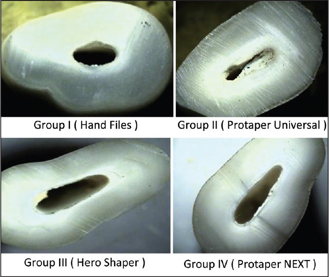
Figure 2.
Stereomicroscopic images showing dentinal defects seen in Groups I, II, III, and IV, showing craze lines seen in Group I. Fracture and other defects in Group II craze lines and partial crack in Group III and craze lines in Group IV
Official websites use .gov
A
.gov website belongs to an official
government organization in the United States.
Secure .gov websites use HTTPS
A lock (
) or https:// means you've safely
connected to the .gov website. Share sensitive
information only on official, secure websites.
Stereomicroscopic images showing dentinal defects seen in Groups I, II, III, and IV, showing craze lines seen in Group I. Fracture and other defects in Group II craze lines and partial crack in Group III and craze lines in Group IV