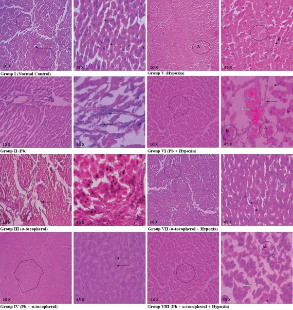
Figure 1.
Hepatic sections of Group I to Group VIII rats: Group I: Hepatic tissue (shown) and hepatic cords (arrows) appeared normal, Group II: Distorted hepatic tissue architecture (circle) and Kupffer cell hyperplasia (arrows), Group III: Thick basement membrane (arrow) and normal regenerating hepatocytes (circle), Group IV: Normal hepatic tissue (shown) and hepatocytes (arrows). Group V: Hepatocytes with swollen mild narrowing sinusoidal spaces (A and B) and foci of necrosis with inflammatory reaction (circle), Group VI: Distorted hepatic parenchyma (circle), foci of fatty change (fine arrows), ballooning degeneration and necrosis of hepatocytes (a), chronic inflammatory cells (b), dilatation and congestion of central vein (block arrow), Group VII: Normal hepatic tissue (circle), little swollen hepatocytes with granular cytoplasm (block arrow), mild narrowing sinusoidal spaces (fine arrows), Group VIII: Hepatic tissue altered lobular architecture (circle), swollen hepatocytes with vacuolated cytoplasm (block arrow), ballooning degeneration (fine arrow), mitotic figure (M)

