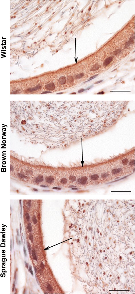
Fig. 3.

Immunolocalization of GPER in the corpus of adult Wistar, Brown Norway and Sprague Dawley rats at higher magnification. GPER immunostaining is observed in the cytoplasm (red color) and immunostaining is strongest near the apical membrane (arrows). Bar = 20 μm
