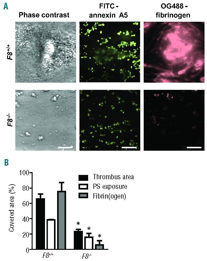
Figure 1.

Role of murine FVIII in the formation of platelet-fibrin thrombi and procoagulant activity in the presence of tissue factor. Blood from wild-type mice or mice deficient in FVIII was perfused over collagen at a rate of shear 1000 s−1 under coagulant conditions. Blood samples were pre-incubated with tissue factor (10 pM) and OG488-fibrinogen. Thrombi were post-stained with FITC-annexin A5 to detect phosphatidylserine (PS) exposure. (A) Representative microscopic phase contrast and fluorescence images after 4 min of flow (bars, 50 μm). (B) Surface area covered by platelet-fibrin thrombi (black bars), PS-exposing platelets (white bars) and fibrin(ogen) (gray bars) as determined by fluorescence studies. Mean ± SEM; n = 8–15; *P<0.05 vs. wild-type.
