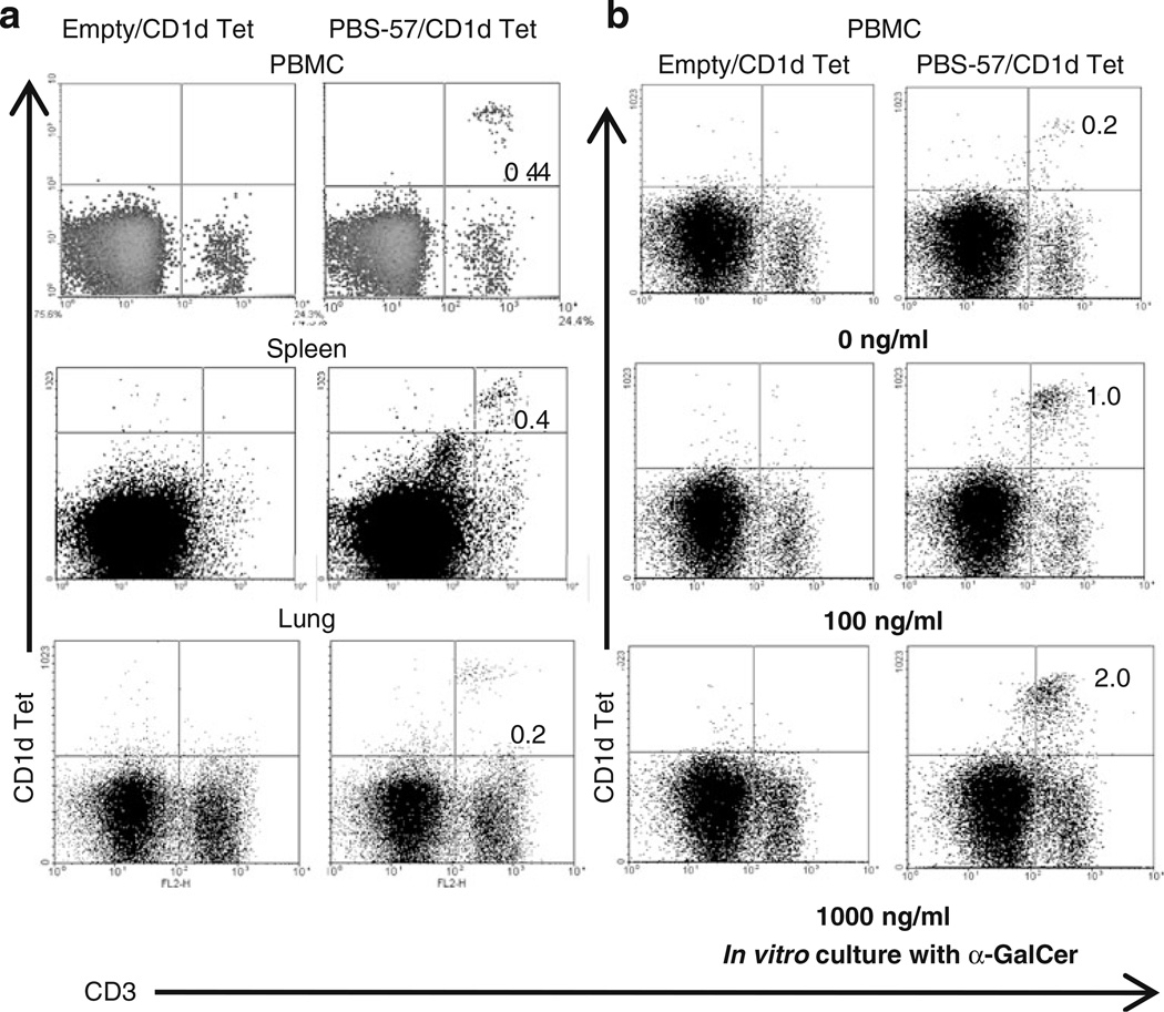
Fig. 2.
iNKT cells are present in pigs. a Two-color flow cytometric analysis of fresh pig PBMCs, splenocytes, and lung MNCs using pig anti-CD3ε mAb conjugated with R-PE and APC-conjugated mouse empty or PBS-57-loaded CD1d tetramers. b Pig PBMCs was cultured in vitro in the presence of various concentrations of α-GalCer for 4 days, and iNKT cells were analyzed as described above. Numbers in the quadrant indicate percentage of cells and the data shown are the representative of five to ten pigs from five independent experiments

