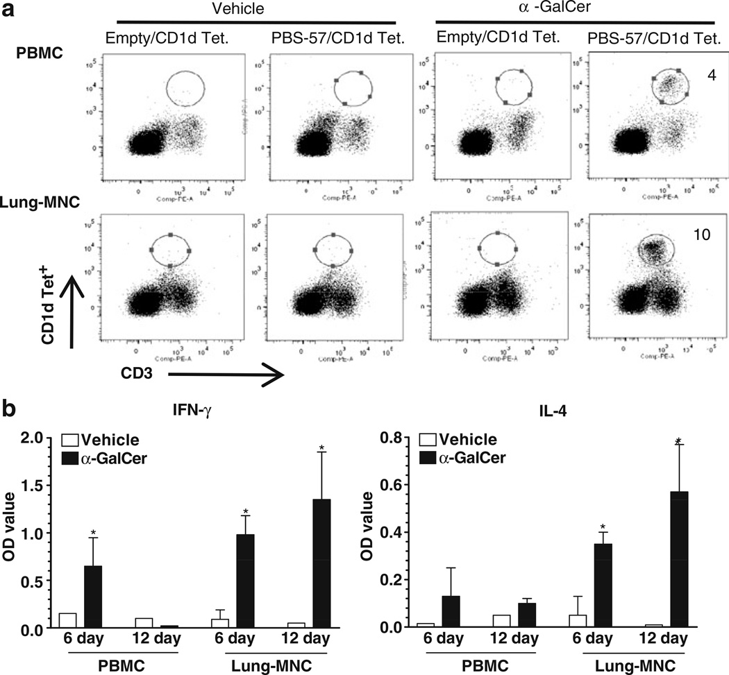
Fig. 3.
Functional iNKT cells are present in pig lungs. Pig lung MNCs and PBMCs (control) were cultured in the presence of α-GalCer (1,000 ng/ml) or vehicle for 12 days. a Immunostained using fluorochrome conjugated anti-pig CD3ε and mouse empty or PBS-57 loaded CD1d tetramers and analyzed by flow cytometry. b Culture supernatants harvested on post-6 and 12 day cultures were analyzed for IFNγ and IL-4 using pig cytokine-specific ELISA. Each bar represents the average cytokines OD from three pigs±SEM and * denote statistically significant difference (P<0.05) in the amount of cytokines secreted by cells cultured in the presence of vehicle vs α-GalCer. The data shown are representative of six pigs in three independent experiments

