Fig. 4.
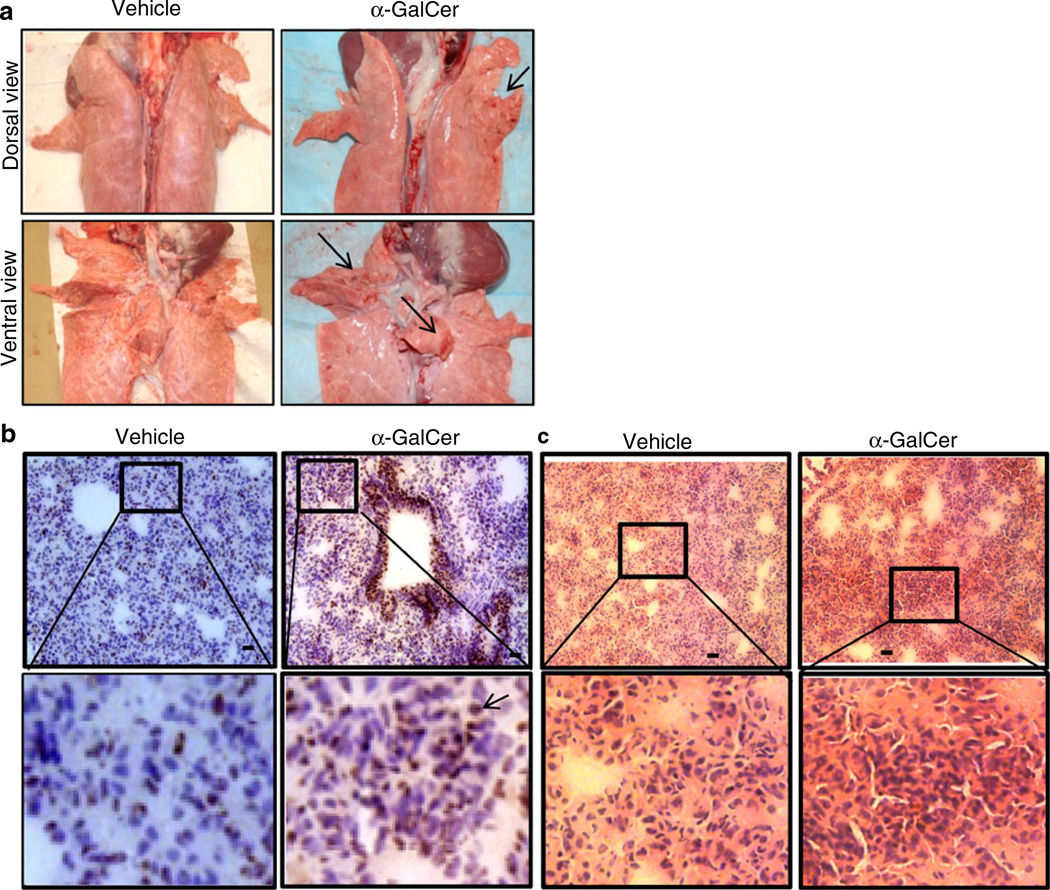
Acute AHR-induced pig lungs had petechial hemorrhages with excessive CD4+ and myeloid cells infiltration. a A representative pig lung macroscopic picture of vehicle or α-GalCer-inoculated pig (n=3 per group) with petechial hemorrhages on both the dorsal and ventral surfaces (arrows indicate areas of hemorrhages) is shown. b A representative pig lung section was subjected to immunohistochemistry, showing more of infiltrated CD4+ cells in α-GalCer received pig lungs compared to vehicle controls. Bar, 10 µm. c A representative pig lung section was subjected to H&E staining, showing massive infiltration of inflammatory leukocytes in α-GalCer received pig lungs. Bar, 5 µm. Frozen sections of vehicle or α-GalCer-inoculated pigs (n=3 per group) were immunostained for pig-specific CD4+ cells (arrowheads) and then counterstained with hematoxylin. Similar results were obtained in another independent experiment

