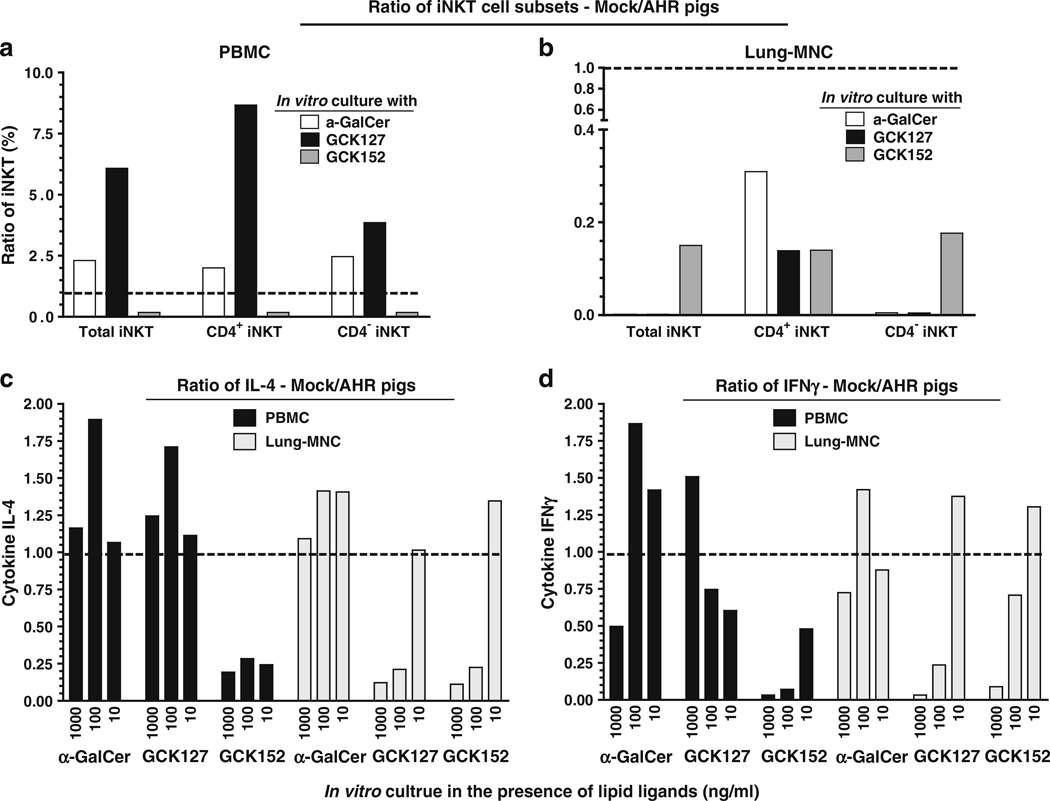
Fig. 6.
Acute AHR-induced pig iNKT cells present in the PBMCs and lung MNCs were preferentially proliferated into CD4+ phenotype and secreted more of IL-4 than IFNγ ex vivo. Pigs were intratracheally inoculated with α-GalCer or vehicle and euthanized at post-24 h of inoculation. a PBMCs and b lung MNCs isolated from pigs were cultured for 8 days in the presence of vehicle, α-GalCer, GCK127, or GCK152 at indicated concentrations. Cells were immunostained using fluorochrome conjugated anti-pig CD3ε, CD4α, and mouse empty or PBS-57-loaded CD1d tetramers and subjected to flow cytometry. CD3+ gated cells were analyzed for mouse CD1d tetramer+ and CD4α± iNKT cells. The supernatants harvested from cultures were analyzed for c IL-4 and d IFNγ by ELISA. Each bar represents the ratio (vehicle/α-GalCer received pigs) of average percentage of iNKT cells or amounts of cytokine from three pigs in one independent experiment. A trend line was drawn on the y-axis at “1”. Ratio of < or > one means that iNKT cells frequency or secreted cytokines were that many fold < or > in AHR-induced pigs compared to respective mock control, respectively. Similar results were observed in another independent experiment

