
Figure 3.
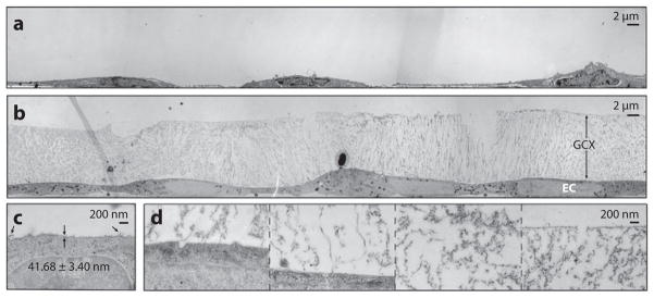
TEM of GCX-covered BAECs (a) preserved conventionally, labeled with ruthenium red and osmium tetroxide, and alcohol dehydrated and (b) preserved by cryo-EM and osmium tetroxide stained. (c) High-magnification image of a conventionally preserved BAEC GCX. Arrows indicate extended strands of GCX. (d) High-magnification image of a cryo-EM BAEC GCX, showing (from left to right) locations near the cell membrane, farther away from the cell membrane, in the center region of the GCX, and at the most apical surface of the GCX. Abbreviations: BAECs, bovine aortic endothelial cells; EM, electron microscopy; GCX, glycocalyx; TEM, transmission electron microscopy. (Figure adapted with permission from 33.)