Fig. 1.

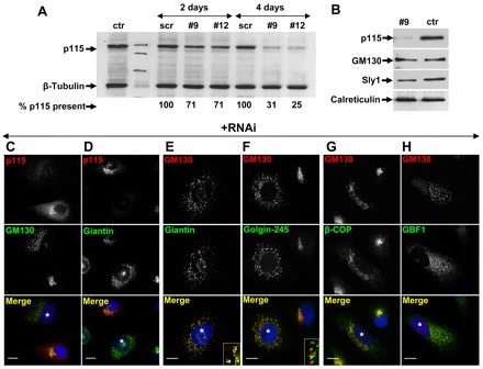
Effects of p115 depletion on Golgi ribbon. (A) HeLa cells mock transfected (ctr), transfected with scrambled (scr) RNA or with siRNA targeting p115 (#9 and #12) were cultured for 2 or 4 days, lysed, and the lysates immunoblotted with the indicated antibodies. Blots were quantified by densitometry to assess p115 levels (numbers below panel). (B) HeLa cell lysates from mock-transfected cells (ctr) or cells silenced with anti-p115 siRNA for 4 days were immunoblotted using the antibodies indicated. (C–H) HeLa cells silenced with anti-p115 siRNA for 4 days were analyzed by immunofluorescence using the antibodies indicated. p115 depletion causes disruption of the Golgi complex (C–D). Golgi fragments show polarized localization of GM130, giantin and golgin-245 (E–F). Insets show higher magnification views. GBF1 and COPI are recruited to Golgi fragments (G–H). p115 depleted cells are marked with *. Scale bars: 10 µm.
