
Fig. 2.
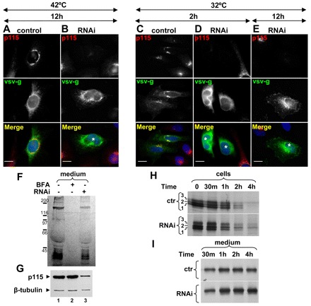
Effect of p115 depletion on cargo traffic. (A–E) Mock-transfected HeLa cells (control) or cells transfected with anti-p115 siRNA for 3 days (RNAi) were transfected with ts045-VSV-G and cultured for additional 12 hours at 42°C (A–B). Cells were shifted to 36°C for indicated times and analyzed by immunofluorescence with the antibodies indicated. In control cells, VSV-G translocates to the Golgi and the plasma membrane (C). In p115-depleted cells, VSV-G is largely retained within the ER (D). In p115-depletd cells after 12 hours at 36°C, VSV-G is detected in internal punctate fragments and the plasma membrane (E). p115-depleted cells are marked with *. Scale bars: 10 µm. (F,G) Untreated cells, cells treated with BFA for 30 minutes and cells silenced with anti-p115 siRNA for 4 days were pulsed with 35S-Met/Cys for 30 minutes and chased with non-radioactive medium (with or without BFA) for 1 hour. Equivalent amounts of cell lysates and media were processed by SDS-PAGE and fluorography. Secretion occurs from control and p115-depleted cells, but not from BFA-treated cells (F). p115 depletion was confirmed by immunoblot of cell lysates with indicated antibodies (G). (H,I) Control cells and cells silenced with anti-p115 siRNA for 3 days were transfected with Myc-tagged cochlin for 24 hours, pulsed with 35S-Met/Cys for 30 minutes and chased with non-radioactive medium for indicated times. At each time point, media were collected and cells were lysed and subjected to immunoprecipitation with anti-Myc. Precipitates were analyzed by SDS-PAGE and fluorography. Cochlin is processed and secreted from control and p115-depleted cells.