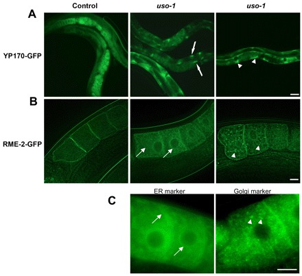
Fig. 3.

Effect of p115 depletion in C. elegans. (A) Localization of YP170–GFP in control and uso-1 siRNA-treated animals. YP170–GFP in RNAi control worms is observed in the intestine, the oocytes and embryos. uso-1-depleted animals show abnormal accumulation of YP170–GFP in the intestine (middle image, arrows) and body cavity (right image, arrowheads). Scale bar: 50 µm. (B) Localization of RME-2–GFP in RNAi control worms and uso-1 siRNA-treated animals. In control worms RME-2–GFP shows predominantly cell surface localization. uso-1-depleted animals show abnormal accumulation of RME-2–GFP in the ER (middle image, arrows) and the Golgi (right image, arrowheads). Scale bar: 10 µm. (C) Localization of the GFP-labeled ER marker protein SP12 and Golgi marker protein UGTP-1 in oocytes. Scale bar: 10 µm.
