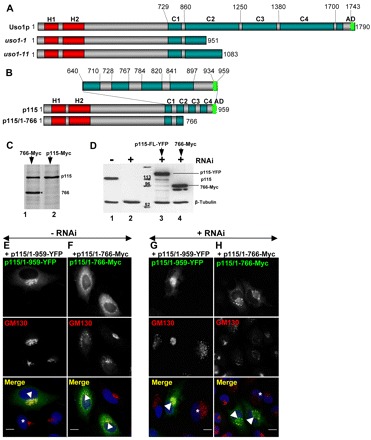
Fig. 4.

p115/1-766 mutant disrupts Golgi ribbon. (A,B) Diagram of full-length Uso1p and Uso1p encoded by uso1-1 and uso1-11, full-length p115 and p115/1-766. H, homology regions between the yeast and mammalian proteins. C, coiled-coil regions. AD, acidic domain. (C) Control HeLa cells (lane 2) or HeLa cells expressing p115/1-766 (lane 1) were labeled with 35S-Met/Cys for 18 hours, lysed and lysates immunoprecipitated with anti-p115 antibodies. Precipitates were processed by SDS-PAGE and fluorography. Similar levels of endogenous p115 and of exogenous p115/1-766 are detected. (D) Control HeLa cells (lane 1) or HeLa cells silenced with anti-p115 siRNA for 3 days (lanes 2–4) were either mock transfected (lane 2), transfected with YFP-p115/1-959 (lane 3) or Myc-p115/1-766 (lane 4) and cultured for additional 18 hours. Cells were lysed and lysates were processed by SDS-PAGE and immunoblotted with indicated antibodies. Cells transfected with anti-p115 RNA oligonucleotides show substantial depletion of endogenous p115 (lane 2). p115-depleted cells transfected with constructs show robust expression of YFP-p115/1-959 (lane 3) or Myc-p115/1-766 (lane 4). (E,F) HeLa cells transfected with GFP-tagged p115/1-959 or p115/1-766 were analyzed by immunofluorescence with indicated antibodies. Expression of p115/1-959 does not alter Golgi morphology (E). Expression of p115/1-766 disrupts Golgi ribbons (F). Scale bars: 10 µm. (G,H) HeLa cells silenced with anti-p115 siRNA for 3 days were transfected with YFP-p115/1-959 or Myc-p115/1-766, cultured for 18 hours and analyzed by immunofluorescence with anti-GM130 and either YFP fluorescence (G) or anti-Myc antibodies (H). Depletion of p115 fragments Golgi ribbon (cells marked with *). Expression of full-length p115 reverses Golgi disruption (G, cell marked with arrowhead). Expression of p115/1-766 does not reverse Golgi disruption (H, cells marked with arrowheads). Scale bars: 10 µm.
