Fig. 7.

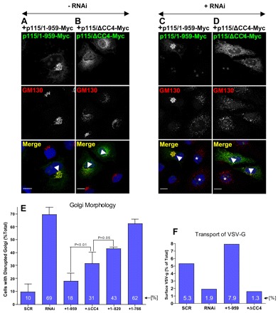
CC4 is important for p115 function. (A,B) HeLa cells transfected with Myc-tagged p115 or p115ΔCC4 for 18 hours were analyzed by immunofluorescence with indicated antibodies. Expression of p115-Myc has no visible effect on Golgi architecture (A, cell marked with arrowhead). Expression of p115ΔCC4-Myc disrupts Golgi ribbon (B, cell marked with arrowhead). Scale bars: 10 µm. (C,D) HeLa cells silenced with anti-p115 siRNA for 3 days were transfected with Myc-tagged p115 or p115ΔCC4 for 18 hours, and analyzed by immunofluorescence with indicated antibodies. p115 depletion fragments the Golgi (cell marked with *). Expression of p115-Myc reverses Golgi disruption (C, cell marked with arrowhead). Expression of p115ΔCC4-Myc does not reverse Golgi disruption (D, cell marked with arrowhead). Scale bars: 10 µm. (E) Golgi disruption was quantified in control cells treated with scrambled RNA (scr), in cells depleted of endogenous p115 (RNAi) and in cells that were depleted of endogenous p115 and expressed either Myc-tagged full-length p115 (+1-959), p115ΔCC4 (+ΔCC4), p115/1-820 (+1-820) or p115/1-766 (+1-766). The values represent the averages of three independent experiments, with more than 50 cells counted each time. (F) VSV-G traffic was quantified in HeLa cells treated with scrambled RNA (scr), depleted of endogenous p115 (RNAi) and in cells that were depleted of endogenous p115 and expressed either Myc-tagged p115/1-959 (+1-959) or p115ΔCC4 (+ΔCC4). The levels of VSV-G present at the plasma membrane after a 2-hour shift to permissive temperature is represented as the percentage of total cellular VSV-G. The values represent the averages of two independent experiments.
